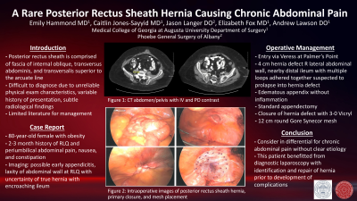
Introduction: Posterior rectus sheath hernias are rare hernias that can be difficult to diagnose due unreliable physical exam characteristics and subtle radiological findings. There is limited literature regarding this topic. As such, we present an interesting case of an elderly female who was found to have a posterior rectus sheath hernia during a diagnostic laparoscopy for chronic abdominal pain.
Case Report: An 80 year-old female with multiple previous abdominal surgeries presented with a 2-3 month history of persistent abdominal pain, constipation, and nausea. CT evaluation revealed possible appendicitis as well as laxity of the abdominal wall of the right lower quadrant with encroaching loops of ileum, but without obvious herniation. She was taken to the operating room for diagnostic laparoscopy and appendectomy. Intraoperatively she was noted to have a 4 cm hernia defect in the right lateral abdominal wall with herniated loops of ileum. An appendectomy and herniorrhaphy with mesh repair were performed. Upon postoperative review of CT imaging and intraoperative photographs, it was determined that this hernia defect is a posterior rectus sheath hernia likely caused by trocar placement from previous laparoscopic surgery.
Discussion and
Conclusion: This report contributes to the limited body of literature that discusses this rare type of hernia and is unique in that both radiological and intraoperative findings are reviewed. Though rare, posterior rectus sheath hernias should be considered in differential diagnoses for patients presenting with chronic abdominal pain without clear etiology. In this case, the patient benefitted from diagnostic laparoscopy as the hernia was identified and repaired prior to developing complications.