Back

Vascular
Category: Quickshot Oral Session 17
Quickshot Oral : Quickshot Oral Session 17
OPTIMAL VISUALIZATION OF THE POPLITEAL ARTERY IN PATIENTS WITH KNEE ARTHROPLASTY.
Monday, February 13, 2023
7:00am – 8:00am East Coast USA Time

- AL
ANA Lozano, DO
Resident physician
Henry Ford Health System, United States - EE
Elisabeth Ekkel, DO
RESIDENT PHYSICIAN
Henry Ford Health System, United States
Presenter(s)
Principal Contact(s)
Objectives: This project was undertaken to examine patient positioning techniques that would allow for better visualization of the popliteal artery in patients with prior knee arthroplasty. Certain position will optimize arteriogram and minimize patient discomfort.
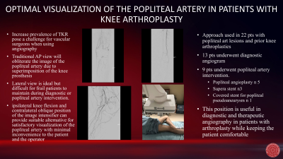
Methods: Following puncture of the common femoral artery (CFA) and completion of the abdominal aortogram and pelvic arteriogram, contralateral CFA is selected. A selective lower extremity arteriogram in anterior/posterior projection is usually performed starting from the groin to the foot. However, visualization of the popliteal artery is inadequate due to overlying metallic knee prosthesis. The knee ipsilateral knee is positioned close to 90 degree lateral flexion and the image intensifier is brought from the opposite side in oblique position of about 25-30 degree popliteal arteriogram.
Results: This approach allows for adequate visualization of the popliteal artery and can be obtained with minimal discomfort to the patient, as lateral knee flexion is better tolerated with the ipsilateral lower extremity almost resting on the xray table. Intervention can also be performed with satisfactory visualization of the popliteal artery with the patient in this position.
Conclusion: The increasing incidence of knee replacement has proven to be a challenge for vascular surgeons when imaging the lower extremity. We have found in our practice that the technique previously discussed has worked well to obtain imaging necessary for diagnosis of vascular pathology and endovascular treatment.

