Back

Vascular
Category: Quickshot Oral Session 17
Quickshot Oral : Quickshot Oral Session 17
SUCCESSFUL COIL EMBOLIZATION OF A LARGE ANTERIOR TIBIAL ARTERY PSEUDOANEURYSM AFTER OPEN REDUCTION INTERNAL FIXATION OF A BICONDYLAR TIBIAL PLATEAU FRACTURE
Monday, February 13, 2023
7:00am – 8:00am East Coast USA Time


Jasmine J. Park, DO
General Surgery Resident
Inspira Health Network
Vineland, New Jersey, United States- SK
Sanjay Kumar, MD
Inspira Health Network, United States
Presenter(s)
Principal Contact(s)
Objectives: Pseudoaneurysms consist of turbulent blood flow between the outside layers of the arterial wall, the tunica media and tunica adventitia. This pathology typically develops after blunt trauma injury to an artery or after catheter-based vascular interventions, but rarely, from arterial injury during orthopedic pinning procedures. Our literature review identified only two cases of tibial artery pseudoaneurysm following closed intermedullary nailing of a proximal tibia fracture.
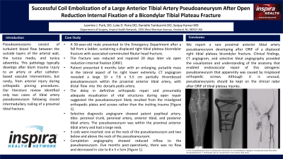
A 56-year-old male presented to the emergency department after a fall, sustaining a displaced right tibial plateau bicondylar fracture with associated comminuted fibular head fracture. The fractures were reduced and repaired 20 days later via open reduction with internal fixation (ORIF). Approximately 6 months later, the patient continued to have discomfort and developed an enlarging pulsatile mass in the lateral aspect of his right lower extremity. A CT angiogram revealed a large 10 x 7.8 x 5.5 cm partially thrombosed pseudoaneurysm within the proximal anterior tibial artery, with distal flow into the dorsalis pedis artery. The delay in definitive orthopedic repair and presumably adequate visualization of vital structures during open repair suggested the pseudoaneurysm likely resulted from the misaligned orthopedic plates and screws rather than the inciting trauma [Figure 1].
A selective diagnostic angiogram revealed a patent popliteal artery, tibio-peroneal trunk, peroneal artery, anterior tibial, and posterior tibial artery. The pseudoaneurysm was within the proximal anterior tibial artery and had a large neck. Subsequently selective angiography and coil embolization inserted 3 coils, one below the neck of the pseudoaneurysm to embolize the anterior tibial artery, and second and third coils placed immediately below and above the neck to isolate the pseudoaneurysm from blood flow. On completion angiography, inflow to the pseudoaneurysm was reduced [Figure 1]. Five months post-embolization an arterial duplex ultrasound revealed no flow within the pseudoaneurysm, which had decreased in size to 8 x 5 x 5 cm [Figure 1].
We report a rare proximal anterior tibial artery pseudoaneurysm developing after ORIF of a displaced right tibial plateau bicondylar fracture. Clinical findings, CT angiogram, and selective tibial angiography provided the visualization and understanding of the anatomy that enabled endovascular repair of this iatrogenic pseudoaneurysm that apparently was caused by misplaced orthopedic screws. Although it is unusual, pseudoaneurysm should be kept on the clinical radar after ORIF of tibial plateau injuries.
Methods:
Results:
Conclusion:

