Introduction: The hepatic inflammatory myofibroblastic tumor (HIMT) is a spindle cell tumor of the liver that originates from the mesenchymal tissues. It is a rare benign tumor with the potential to degenerate into a malignant and invasive tumor. It can occur anywhere in the body with the most common sites being the lung, mesentery, and omentum. We present a rare case of a HIMT in an elderly female with active primary squamous lung cancer (SCC).
Case Presentation: An 81-year-old woman with abdominal discomfort was found to have a nonspecific mass within the dome of the liver measuring 2.2cm on Computed tomography (CT). Upper and lower gastrointestinal endoscopies were negative for any gastrointestinal malignancy. A metastatic workup revealed a 1.8cm solitary lung nodule concerning for primary lung cancer. She underwent thoracoscopic wedge resection of the lung nodule with a core needle biopsy of the liver mass. The pathology of the resected lung nodule indicated the mass to be primary keratinizing squamous cell carcinoma, while the liver biopsy indicated the liver mass to be a primary hepatic inflammatory myofibroblastic tumor. Six months after her lung surgery, she underwent non-anatomical resection of the liver tumor including hepatic segments VII and VIII with negative margins on histological analysis. She remained disease-free for four years when she had a metastatic recurrence of HIMT measuring 5.8cm by 2.9cm extending to the chest wall. She declined any further treatment for her metastatic disease.
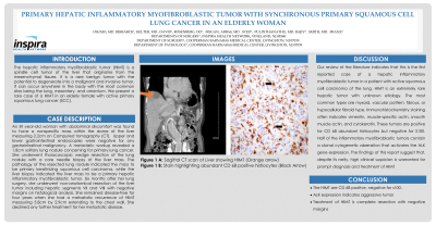
Discussion/conclusion Our review of the literature indicates that this is the first reported case of a hepatic inflammatory myofibroblastic tumor in a patient with active squamous cell carcinoma of the lung. HIMT is an extremely rare hepatic tumor with unknown etiology. The most common types are myxoid, vascular pattern, fibrous, or hypocellular fibroid type. Immunohistochemistry staining often indicates vimentin, muscle-specific actin, smooth muscle actin, and cytokeratin. These tumors are positive for CD 68 abundant histiocytes but negative for S100. Half of the inflammatory myofibroblastic tumors contain a clonal cytogenetic aberration that activates the ALK gene expression. The findings of this report suggest that, despite its rarity, high clinical suspicion is warranted for prompt diagnosis and treatment of HIMT.